Knowledge Center · DIGITAL DENTISTRY
Intraoral Scanning Is Not Just “Taking Pictures of Teeth”
Intraoral scanning is not just a replacement for traditional impressions. Its real value is creating 3D records that allow treatment stages, design meshes, final restorations, orthodontic outcomes, and long-term maintenance to be compared over time.
Many patients hear “intraoral scanning” and think it simply means using a device to take a few pictures inside the mouth.
In digital dentistry, however, intraoral scanning is not a regular photograph, and it is not only a replacement for traditional impressions. Its more important value is that it records teeth, gums, bite relationships, and treatment stages as 3D data, allowing dentists to compare, overlay, and track changes across different time points.
In other words, an intraoral scan does not only record “what the mouth looks like now.” It helps make the treatment process something that can be saved, reviewed, and verified.
An intraoral scan records a 3D mesh, not a photograph
An intraoral scan converts tooth shape, gingival margins, arch relationships, and bite information into a 3D model. In a digital workflow, this model can be understood as a mesh: a digital structure made of many three-dimensional points and surfaces.
This mesh is not just a visual model for patients to look at. It can be measured, designed on, compared, and used for fabrication by dentists, technicians, and digital design software.
For veneers, ceramic crowns, implant restorations, orthodontics, bite analysis, DSD smile design, and long-term maintenance, scan data can become part of the clinical decision-making foundation.
The key point is that every scan can become a record of a specific moment in treatment.
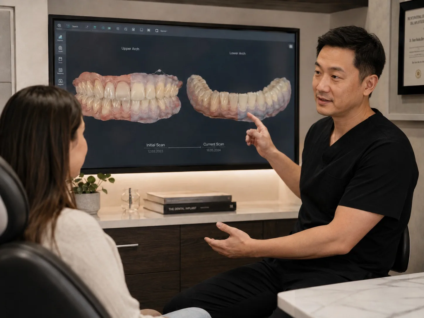
The strongest value of scanning is stage-by-stage comparison
Traditional models can also record the oral condition, but comparing multiple treatment stages quickly and precisely is much harder. With intraoral scanning, 3D models from different time points can be stored and overlaid in software.
For example:
- The initial scan can record the condition before treatment.
- The Mockup scan can record how the proposed design actually appears in the mouth.
- The provisional restoration scan can record the transition stage.
- The final restoration scan can record the completed result.
- Maintenance scans can record whether teeth, gums, or restorations have changed over time.
When these records are overlaid, dentists do not need to rely only on the impression that “something seems different.” They can more clearly see where the change occurred, how much change occurred, and whether it is within an acceptable range.
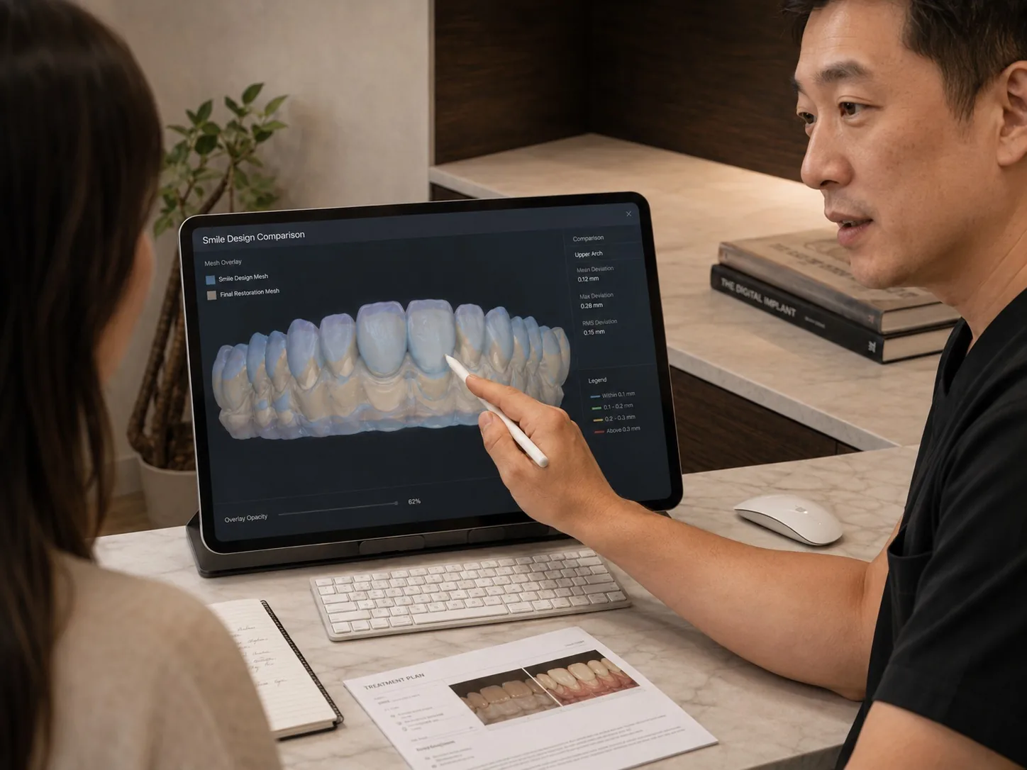
The design mesh and final restoration can be compared
In digital aesthetic dentistry, a proposed treatment design can be converted into a 3D design mesh, not just a flat image.
After treatment is completed, the final restoration can also be captured with an intraoral scan as an actual mesh. Overlaying the design mesh and the final restoration mesh can help the dentist evaluate:
- Whether the final tooth shape is close to the original design.
- Whether the length, thickness, and proportions meet the intended plan.
- Whether symmetry and the smile line remain consistent.
- Whether restoration contours or margins need adjustment.
- Where the real intraoral result differs from the proposed design.
This is one of the important values of intraoral scanning in DSD and aesthetic restoration. It is not only used to “make teeth.” It connects design, Mockup, provisional restorations, and final restorations with clearer clinical verification.
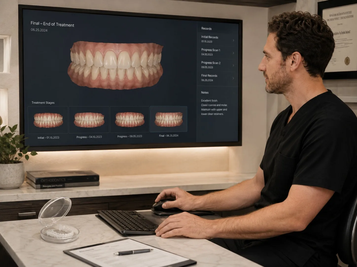
Orthodontic treatment also benefits from staged mesh records
In orthodontics, intraoral scans can record tooth positions at different stages of treatment. Each scan can become a digital record of that stage and can be compared with the initial model, an interim target, or the final goal.
This helps the dentist evaluate whether tooth movement is following the plan, whether the bite is becoming stable, whether the arch form has changed, and whether additional refinement is needed.
For patients, these scan comparisons can also make treatment progress easier to understand. Tooth movement is not always obvious day to day, but stage-by-stage mesh comparison can show changes in tooth position and arch form more clearly.
After orthodontic treatment, the final scan can also be used to fabricate retainers and serve as a baseline for future reviews. If mild tooth movement occurs years later, a new scan can be overlaid with the final orthodontic scan to identify where the change happened.
Intraoral scanning helps preserve long-term tooth form
Many dental treatments do not end on the day they are completed. Long-term maintenance is the real test.
Intraoral scanning can help preserve tooth form at a specific stage, such as:
- The final shape after veneers or ceramic crowns.
- The tooth position after orthodontic treatment.
- The bite relationship after implant restoration.
- The occlusal surface condition in patients with bruxism.
- The gingival and arch changes in periodontal maintenance.
These records can become important references for future follow-up, repair, remaking, or retainer fabrication.
If a restoration fractures years later, a retainer is lost, or teeth have shifted, the previous scan data is not just an old file. It can help the dentist understand the original condition more quickly and reduce guesswork and repeated communication.
Retainers, night guards, and remake work all benefit
One practical value of scanning in long-term maintenance is that digital records can be saved and reused when appropriate.
After orthodontic treatment, a retainer can be fabricated from the final scan.
If a retainer is lost, the existing digital record may help when a replacement is clinically appropriate.
For patients who grind their teeth, scans taken at different times can help observe wear or changes in the bite surface.
When a restoration needs repair or replacement, the previous digital model can help the dentist and technician understand the original shape, bite, and space.
Patients may not feel this value immediately during the first appointment, but it becomes very important in long-term maintenance. It turns dental treatment from a one-time intervention into a process that can be tracked and managed digitally.
How is this different from traditional impressions?
Traditional impressions can create physical models and still have value in many clinical situations. But physical models are harder to store, duplicate, transmit, and compare across multiple stages.
The advantage of intraoral scanning is not simply that it is “faster.” The data can be digitally stored, copied, transmitted, overlaid, and compared.
This makes it easier for dentists and technicians to communicate using the same data foundation, and it makes changes between treatment stages easier to see.
Of course, scanning is not magic. Accuracy is still affected by the device, intraoral conditions, scan path, soft tissue, saliva, reflective materials, and operator technique. Digital dentistry is not meant to replace clinical judgment. It is meant to give clinical judgment a stronger data foundation.
How D4 uses intraoral scanning
At D4, intraoral scanning is not used to show off a device. It is used to build a more complete digital treatment record.
The questions we care about are: What problem does this scan need to solve? Which other records should it connect with? Will it need to be compared again in the future?
For aesthetic restoration, intraoral scans can connect DSD design, Mockup, provisional restoration, and final restoration.
For orthodontics, scans can record treatment stages and the final tooth position.
For implant restoration, scans can be combined with CBCT to plan the implant position from the future crown position backward.
For long-term maintenance, scans can preserve the completed shape as a reference for future follow-up, retainers, night guards, or restoration maintenance.
So intraoral scanning is not just “taking pictures of teeth.”
It records a patient’s 3D oral condition across different stages of treatment.
The value of digital dentistry is not only turning records into computer files. It is making treatment something that can be designed, compared, verified, and preserved over time.
FAQ
What is intraoral scanning?
Intraoral scanning records the teeth, gums, and bite relationship with a digital scanner and turns them into a 3D model. This model is not a regular photo. It can be used for diagnosis, design, restoration fabrication, orthodontic evaluation, implant planning, and long-term maintenance.
How is intraoral scanning different from traditional impressions?
Traditional impressions mainly create a physical model, while intraoral scanning creates a digital 3D model. The advantage is not only speed. Digital models can be stored, copied, transmitted, overlaid, and compared. For veneers, crowns, orthodontics, implant restorations, and maintenance, this makes communication and tracking between treatment stages more efficient.
Can intraoral scans compare changes before and after treatment?
Yes. One important value of intraoral scanning is that it can record 3D models from different treatment stages and allow them to be overlaid and compared. Initial records, Mockup scans, provisional restoration scans, final restoration scans, and maintenance scans can all become time-point records for evaluating changes in tooth shape, position, bite, and restoration outcomes.
Can the final veneer or smile restoration be compared with the original design?
Yes. In digital aesthetic dentistry, the proposed design can exist as a design mesh, and the final restoration can be captured as an actual scan mesh. Overlaying the two helps the dentist evaluate whether the final tooth shape, length, thickness, proportions, and symmetry are close to the design goal. This is an important part of DSD and veneer treatment verification.
Why should orthodontic scan records be kept after treatment?
The final scan after orthodontic treatment records the completed tooth position and can be used to fabricate retainers. If a retainer is lost, if mild tooth movement occurs, or if relapse needs to be evaluated, a new scan can be compared with the final orthodontic scan. This is clearer than relying on visual judgment alone and supports long-term maintenance.
Are scan records useful for remaking retainers, night guards, or restorations?
Yes. Intraoral scan records preserve tooth shape, bite relationship, and restoration outcomes from a specific stage. If a retainer is lost, a night guard needs to be remade, or a restoration needs repair or replacement, previous digital models can be important references. They do not replace a new clinical examination, but they can reduce guesswork and repeated communication.